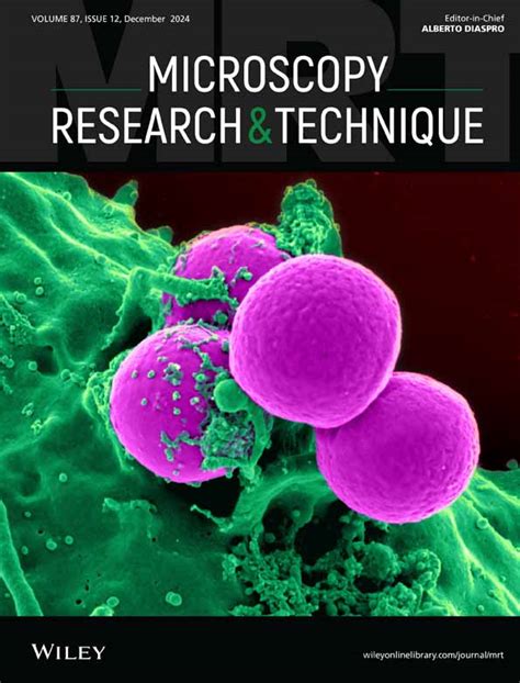
The world of microscopy research and techniques has revolutionized our understanding of the microscopic world, enabling us to explore and study the intricacies of cells, microorganisms, and materials at an unprecedented level of detail. From the early days of microscopy to the present, this field has undergone significant advancements, transforming the way we conduct research, diagnose diseases, and develop new technologies.
The Evolution of Microscopy Techniques

The history of microscopy dates back to the 17th century when Antonie van Leeuwenhoek first observed microorganisms using a simple microscope. Since then, microscopy techniques have undergone significant transformations, from the development of light microscopes to the invention of electron microscopes, and more recently, the emergence of advanced techniques such as super-resolution microscopy and live-cell imaging.
Advancements in Microscopy Techniques
The development of new microscopy techniques has enabled researchers to study biological samples with unprecedented resolution and sensitivity. Some of the key advancements in microscopy techniques include:
- Super-resolution microscopy: This technique allows researchers to study biological samples at the nanoscale, enabling the visualization of individual molecules and their interactions.
- Live-cell imaging: This technique enables researchers to study living cells in real-time, allowing for the observation of cellular dynamics and behavior.
- Electron microscopy: This technique uses a beam of electrons to produce high-resolution images of biological samples, enabling the study of cellular structures and organelles.
The Impact of Microscopy Research on Medicine

Microscopy research has had a significant impact on medicine, enabling the diagnosis and treatment of diseases at the cellular and molecular level. Some of the key ways in which microscopy research has impacted medicine include:
- Cancer research: Microscopy techniques have enabled researchers to study the behavior of cancer cells, leading to a better understanding of the disease and the development of new treatments.
- Infectious diseases: Microscopy techniques have enabled researchers to study the behavior of microorganisms, leading to a better understanding of the diseases they cause and the development of new treatments.
- Regenerative medicine: Microscopy techniques have enabled researchers to study the behavior of stem cells, leading to a better understanding of their role in tissue repair and regeneration.
The Role of Microscopy in Disease Diagnosis
Microscopy techniques play a critical role in disease diagnosis, enabling healthcare professionals to visualize and study biological samples at the cellular and molecular level. Some of the key ways in which microscopy is used in disease diagnosis include:
- Histopathology: Microscopy techniques are used to study tissue samples, enabling the diagnosis of diseases such as cancer.
- Cytology: Microscopy techniques are used to study cell samples, enabling the diagnosis of diseases such as infections.
- Microbiological diagnosis: Microscopy techniques are used to study microorganisms, enabling the diagnosis of diseases such as tuberculosis.
The Future of Microscopy Research and Techniques

The future of microscopy research and techniques holds much promise, with the potential to revolutionize our understanding of the microscopic world and drive innovation in fields such as medicine, materials science, and biotechnology. Some of the key areas of research and development in microscopy include:
- Advanced imaging techniques: Researchers are developing new imaging techniques such as super-resolution microscopy and live-cell imaging, enabling the study of biological samples with unprecedented resolution and sensitivity.
- Artificial intelligence and machine learning: Researchers are applying artificial intelligence and machine learning algorithms to microscopy data, enabling the automated analysis of large datasets and the discovery of new insights.
- Integration with other technologies: Researchers are integrating microscopy techniques with other technologies such as spectroscopy and microfluidics, enabling the study of biological samples in new and innovative ways.
Challenges and Opportunities in Microscopy Research
Despite the many advances in microscopy research and techniques, there are still many challenges and opportunities in this field. Some of the key challenges and opportunities include:
- Data analysis and interpretation: The large amounts of data generated by microscopy techniques require advanced computational tools and expertise to analyze and interpret.
- Standardization and validation: Microscopy techniques require standardization and validation to ensure that results are reliable and reproducible.
- Access and affordability: Microscopy techniques and equipment can be expensive, limiting access to researchers and healthcare professionals in resource-poor settings.
Gallery of Microscopy Techniques


We invite you to share your thoughts on the impact of microscopy research and techniques on medicine and other fields. How do you think advancements in microscopy will shape the future of research and innovation? Share your comments and questions below.
What is microscopy research?
+Microscopy research refers to the use of microscopy techniques to study biological samples at the cellular and molecular level.
What are some common microscopy techniques?
+Some common microscopy techniques include super-resolution microscopy, live-cell imaging, electron microscopy, fluorescence microscopy, confocal microscopy, and two-photon microscopy.
How has microscopy research impacted medicine?
+Microscopy research has had a significant impact on medicine, enabling the diagnosis and treatment of diseases at the cellular and molecular level.
