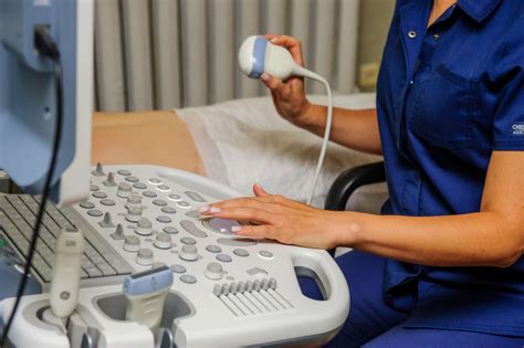
The field of ultrasound technology has revolutionized the way medical professionals diagnose and treat various health conditions. Ultrasound technicians, also known as sonographers, play a crucial role in capturing high-quality images of internal organs and tissues, which enable doctors to make accurate diagnoses and develop effective treatment plans. In this article, we will explore the ways ultrasound techs take amazing diagnostic pictures.
The importance of ultrasound technology cannot be overstated. It is a non-invasive, pain-free, and relatively low-cost imaging modality that provides real-time images of internal structures. Ultrasound techs use specialized equipment to produce high-frequency sound waves that penetrate the body, bounce off internal structures, and return to the transducer as echoes. These echoes are then converted into images that can be interpreted by medical professionals.
1. Understanding the Fundamentals of Ultrasound Technology
To take amazing diagnostic pictures, ultrasound techs must have a solid understanding of the fundamentals of ultrasound technology. This includes knowledge of the physical principles of sound waves, the characteristics of different tissues, and the equipment used to produce and display images. Ultrasound techs must also be familiar with the various modes of ultrasound imaging, including 2D, 3D, and Doppler.

Key Principles of Ultrasound Imaging
- Frequency: Ultrasound imaging uses high-frequency sound waves, typically in the range of 2-15 MHz.
- Wavelength: The wavelength of the sound wave determines the resolution of the image.
- Tissue interaction: Ultrasound waves interact with tissues in different ways, depending on their composition and density.
2. Choosing the Right Equipment and Settings
Ultrasound techs must choose the right equipment and settings to produce high-quality images. This includes selecting the appropriate transducer, adjusting the frequency and gain, and optimizing the image processing settings. The type of transducer used depends on the body part being imaged and the specific clinical question being addressed.

Factors Affecting Image Quality
- Transducer selection: The type of transducer used affects the frequency and resolution of the image.
- Frequency adjustment: The frequency of the sound wave affects the depth of penetration and the resolution of the image.
- Gain adjustment: The gain setting affects the brightness of the image.
3. Preparing the Patient for the Procedure
Ultrasound techs must prepare the patient for the procedure by explaining the process, positioning the patient correctly, and ensuring that the skin is clean and free of lotions or oils. This helps to ensure that the images are of high quality and that the patient is comfortable during the procedure.

Tips for Optimizing Patient Positioning
- Use a comfortable and stable position to minimize movement artifacts.
- Use pillows or wedges to support the patient's body and maintain the desired position.
- Ensure that the skin is clean and free of lotions or oils to improve transducer contact.
4. Capturing High-Quality Images
Ultrasound techs must capture high-quality images by adjusting the transducer position, angulation, and pressure to optimize the image. This may involve using different scanning techniques, such as sector scanning or linear scanning, to capture the desired image.

Tips for Optimizing Image Quality
- Use the correct scanning technique for the specific body part being imaged.
- Adjust the transducer position, angulation, and pressure to optimize the image.
- Use the correct frequency and gain settings to optimize image quality.
5. Analyzing and Interpreting Images
Finally, ultrasound techs must analyze and interpret the images to ensure that they are of high quality and provide the necessary diagnostic information. This involves evaluating the image for artifacts, measuring distances and velocities, and identifying normal and abnormal structures.

Key Components of Image Analysis
- Image evaluation: Evaluate the image for artifacts, resolution, and contrast.
- Measurement: Measure distances and velocities to provide quantitative data.
- Identification: Identify normal and abnormal structures to provide diagnostic information.
Gallery of Ultrasound Imaging Techniques


In conclusion, ultrasound techs play a critical role in capturing high-quality images that enable doctors to make accurate diagnoses and develop effective treatment plans. By understanding the fundamentals of ultrasound technology, choosing the right equipment and settings, preparing the patient for the procedure, capturing high-quality images, and analyzing and interpreting images, ultrasound techs can produce amazing diagnostic pictures that improve patient care.
We invite you to share your thoughts and experiences on ultrasound technology in the comments section below. Have you had a positive experience with ultrasound imaging? Do you have any questions or concerns about the technology? Share your stories and let's start a conversation!
What is ultrasound technology?
+Ultrasound technology is a medical imaging modality that uses high-frequency sound waves to produce images of internal organs and tissues.
What is the role of an ultrasound tech?
+An ultrasound tech, also known as a sonographer, is a medical professional who uses ultrasound technology to capture high-quality images of internal organs and tissues.
What are the benefits of ultrasound technology?
+Ultrasound technology is non-invasive, pain-free, and relatively low-cost, making it a valuable diagnostic tool in medical imaging.
